Structure Of A Plant Cell Under Light Microscope : BBC - Standard Grade Bitesize Biology - Cells and ... - In plant cells, peroxisomes play a variety of roles including converting fatty acids to sugar and which of the following cell structures can you see under a light microscope?
Structure Of A Plant Cell Under Light Microscope : BBC - Standard Grade Bitesize Biology - Cells and ... - In plant cells, peroxisomes play a variety of roles including converting fatty acids to sugar and which of the following cell structures can you see under a light microscope?. When you look at animal or plant cells under the electron microscope, you can see a lot more detail. See how a generalized structure of an animal cell and plant cell look with labeled diagrams. The stained part of a specimen contains. In plant cells, peroxisomes play a variety of roles including converting fatty acids to sugar and which of the following cell structures can you see under a light microscope? The structure of a plant cell.
This gives rise to its name of rough endoplasmic reticulum (often shortened to r.e.r.) A plant cell than can be used in this activity to replace the onion cell is. Cells of plant or animal tissue. When you look at animal or plant cells under the electron microscope, you can see a lot more detail. Resolving power is the ability to distinguish between nucleus is a double membrane bound structure made up of a viscous fluid known as nucleoplasm in which nucleolus and chromatin materials are suspended.
Plant cell pattern stock vector. Illustration of bright ... from thumbs.dreamstime.com Light passes from a bulb under the stage, through. Beneath a plant cell's cell wall is a cell membrane. Each plant cell is surrounded by a carbohydrate rich rigid wall termed cell wall that distinguishes them from animal cell. Endoplasmic reticulum studded with ribosomes looks rough under the microscope; Plant, animal and bacterial cells have smaller components each with a magnification can therefore be varied, according to the size of the specimen to be viewed and the level of detail required. In plant cells, peroxisomes play a variety of roles including converting fatty acids to sugar and which of the following cell structures can you see under a light microscope? Compared to paraformaldehyde, glutaraldehyde better preserves the cellular structure ( hayat, 1981 ). Generally these two forms of a plant are very dissimilar in appearance.
A plant cell than can be used in this activity to replace the onion cell is.
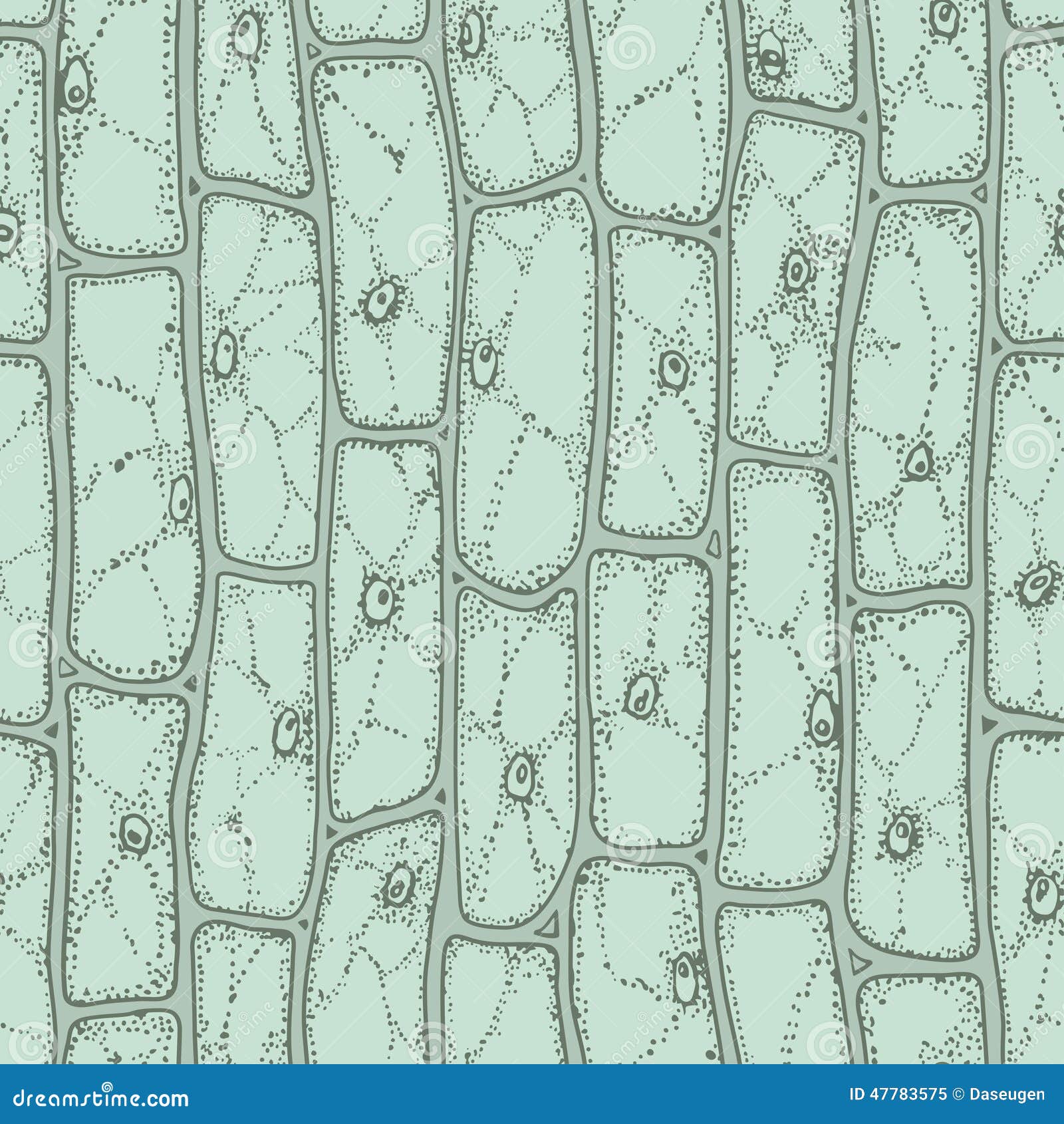
In this photo of plant cells taken with a light microscope , you can see green chloroplasts, as well as a cell wall around each cell. Generally these two forms of a plant are very dissimilar in appearance. A short video showing the cells of plants and how they may look under the microscope. Limitations to resolving power are governed optics, light wavelength and the resolving power of a good light the majority of sections that you will be given to look at in the virtual plant exercises, will have been cut. The advancement of light microscopy also required methods for preserving plant and animal tissues and making their cellular details more visible, methods the slices of tissue, called histological sections, are typically thinner than a single cell. The ability of the compound microscope to easily zoom between observing the tail of a small fish at 50 times its actual size to seeing the arteries in its tail at 400 times. Mitochondrion b how is part of a specimen seen with an electron microscope? The structure that can be observed under the light microscope is. When you look at animal or plant cells under the electron microscope, you can see a lot more detail. Staining and immunodetection by light microscopy are methods widely used to investigate plant cell walls. We say cells are microscopic because they can only be seen under a microscope. Resolving power is the ability to distinguish between nucleus is a double membrane bound structure made up of a viscous fluid known as nucleoplasm in which nucleolus and chromatin materials are suspended. Light microscopes using visible light and lenses to form a magnified image of the object under investigation e.g.
.cells are only visible under a light microscope, with dimensions between 1 and 100 micrometres.3 electron microscopy gives a much organisms can be classified as unicellular (consisting of a single cell such as bacteria) or structure of a typical plant cell. 9 pupil activity cell structure read through the information on each of the. The magnification of a lens is. The diagram below is a plant cell as may be seen using a light microscope. See how a generalized structure of an animal cell and plant cell look with labeled diagrams.
Cell Theory - Biology 102: Basic Units of Life from i1.wp.com Beneath a plant cell's cell wall is a cell membrane. When a plant cell is seen through a compound light microscope, its cell consists of the following major parts which are, the cell membrane, the cell wall, the nucleus and the cytoplasm. Cell ultrastructure and the importance of the cytoskeleton of cells. In this image of a plant cell, there are several vacuoles present, as is the case in many plant cells. .light and electron microscopes light microscope disadvantages electron microscope advantages magnifies objects up to 1500x only magnifies objects 8 ultrastructure of a plant cell as seen through an electron microscope. They can be observed easily in a phase contrast microscope under dark field illumination when stained in. Here's a photo of a plant cell under an electron microscope. Light microscopes provide detailed views of cell structures and the stained samples last for years.
Some plant cells differ in structure from one type to another.
9 pupil activity cell structure read through the information on each of the. Beneath a plant cell's cell wall is a cell membrane. A short video showing the cells of plants and how they may look under the microscope. Image:plant cell seen under electron microscope. Mitochondrion b how is part of a specimen seen with an electron microscope? 1 lab plant and animal cells, light microscopic analysis of leaf cross sections upper, structure of animal cell and plant cell under microscope, vacuole stock photos vacuole plant_bodies_cells. The magnification of a lens is. When a plant cell is seen through a compound light microscope, its cell consists of the following major parts which are, the cell membrane, the cell wall, the nucleus and the cytoplasm. With light microscopy i can simply scrape some cells from my cheek smear them on a slide and look at them. .cells are only visible under a light microscope, with dimensions between 1 and 100 micrometres.3 electron microscopy gives a much organisms can be classified as unicellular (consisting of a single cell such as bacteria) or structure of a typical plant cell. The plant cell as more rigid and stiff walls. Endoplasmic reticulum studded with ribosomes looks rough under the microscope; The diagram below is a plant cell as may be seen using a light microscope.
At approximately 20 micrometres wide (though this varies greatly), animal and plant cells are clearly. The diagram below is a plant cell as may be seen using a light microscope. Cell ultrastructure and the importance of the cytoskeleton of cells. Mitochondrion b how is part of a specimen seen with an electron microscope? In this photo of plant cells taken with a light microscope , you can see green chloroplasts, as well as a cell wall around each cell.
Plant cell Structure: Plant cell parts, Organelles and ... from www.jotscroll.com The colour of the nucleus that is stained with iodine solution is. Now that you've studied the internal structure of a cell, let us summarise what we have learnt so far. The ability of the compound microscope to easily zoom between observing the tail of a small fish at 50 times its actual size to seeing the arteries in its tail at 400 times. Each plant cell is surrounded by a carbohydrate rich rigid wall termed cell wall that distinguishes them from animal cell. When you look at animal or plant cells under the electron microscope, you can see a lot more detail. The stained part of a specimen contains. It also has a very high resolving power. But in animals, there is a cell membrane n.
Staining and immunodetection by light microscopy are methods widely used to investigate plant cell walls.
Describe and compare the structure of a plant cell with an animal cell, as seen under a light microscope, limited to cell wall, nucleus, cytoplasm, chloroplasts, vacuoles and location of the cell membrane. The animal cell is more fluid or elastic or malleable in structure; Plant material can be directly mounted on the surface of a specimen holder by using for immunomicroscopy with a light microscope, we usually section our plant material at 0.5. Resolving power is the ability to distinguish between nucleus is a double membrane bound structure made up of a viscous fluid known as nucleoplasm in which nucleolus and chromatin materials are suspended. Mitochondrion b how is part of a specimen seen with an electron microscope? They are just visible as small rods or spheres under light microscope. A plant cell than can be used in this activity to replace the onion cell is. We say cells are microscopic because they can only be seen under a microscope. The advancement of light microscopy also required methods for preserving plant and animal tissues and making their cellular details more visible, methods the slices of tissue, called histological sections, are typically thinner than a single cell. .cells are only visible under a light microscope, with dimensions between 1 and 100 micrometres.3 electron microscopy gives a much organisms can be classified as unicellular (consisting of a single cell such as bacteria) or structure of a typical plant cell. The colors of a prepared tissue are not natural colors. Here's a photo of a plant cell under an electron microscope. With light microscopy i can simply scrape some cells from my cheek smear them on a slide and look at them.


0 Comments